


Visualizing the invisible with unprecedented resolution
with Prof. Yasukazu Murakami


Many times stronger than the magnets many of us have on our white boards or refrigerators, high-performance permanent magnets are enabling efficient motors and generators in technologies such as electric cars and wind turbines to help society move away from fossil fuels and toward cleaner and renewable energy sources.
However, despite their name, permanent magnets can lose their magnetization at elevated temperatures. Thus, scientists are looking for any edge to make the magnets stronger and more resistant to thermal demagnetization in the pursuit of better efficiency for a sustainable society.
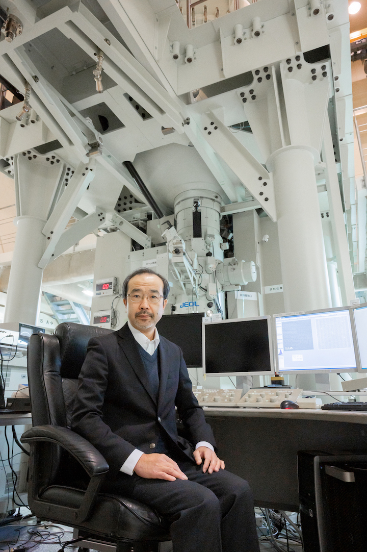
Yasukazu Murakami, professor of Kyushu University’s Faculty of Engineering, is approaching this issue on the nanometer scale using the university’s extremely powerful electron microscopes. But instead of looking at just the physical structures of the magnetics, he is visualizing the magnetic fields themselves.
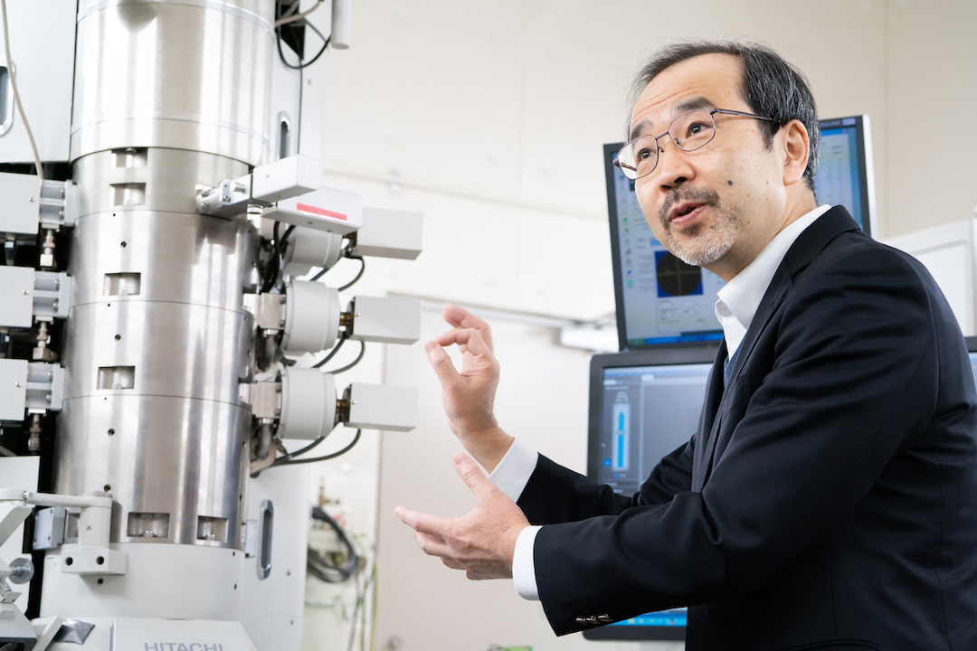
“While electron microscopes are known for producing high-resolution images that can allow us to observe the very arrangement of atoms in a material, the beam of electrons used to image samples is also influenced by magnetic and electric fields,” explains Murakami.
“Through electron holography, we can get information about these magnetic and electric fields by essentially comparing a beam of electrons affected by the fields with a reference beam that hasn’t undergone these effects.”
Murakami and his group have been working to improve the technique and observe the fields with pin-point precision in the highest resolution possible. Though permanent magnetics used in cars and wind turbines are by no means small, further improvements depend on understanding them at the tiniest level.
“Commonly used permanent magnetics may look like a continuous, single material to the human eye, but they are actually composed of tiny crystal grains. In addition to adjacent grains having different compositions and orientations, boundaries between grains are hot spots of variations that can affect overall performance,” says Murakami.
Until recently, grain boundaries were thought to have a composition that would make them non-magnetic, but recent work showed this not to be the case. Murakami and his group has been focusing their microscopes on these grain boundaries to measure the magnetism at these specific points and better understand how they contribute to demagnetization when operated at extremes.
Electron holography’s applications are not limited to just magnetism. The technique can also be used to probe electric fields—which are critical for everything from operation of electronic devices to the progress of chemical reactions—on microscope scales.
Just last October, publishing in the journal Science, Murakami’s group and his collaborators were able to count the number of extra—or missing—charges down to a precision of just one electron in single platinum catalyst nanoparticles with diameters of only 10 nm, one-tenth the size of common viruses.
Differences of just a single electron can have a profound impact on how metal nanoparticle catalysts facilitate reactions such as the breakdown of greenhouse and other harmful gases into benign ones and the efficient production ammonia for fertilizers used in agriculture. Thus, this new level of understanding will aid in developing ways to achieve a sustainable future.

To make these observations possible, Murakami and his group are developing and applying new ways to push resolution to new limits, such as by using sophisticated optics for electrons interference, employing high-sensitivity camera, using machine learning to improve the accuracy of detecting particles for measurement, and utilizing new models to reduce noise in data.
Each step helps to unlock the fundamental workings behind various processes and phenomenon in more detail, potentially leading to breakthroughs in a wide range of fields.
“Microscopes have shown us cells and then atoms. Now, through the creativity of scientists, we are visualizing the invisible on extremely tiny scales, which allows us to understand physics on a new level and verify theories,” says Murakami. “Our continued efforts probing the microscale will help drive development of technologies for a variety of macroscale applications in our everyday lives.”