![]()


![]()
![]()
研究成果 Research Results
- TOP
- News
- Research Results
- Pathological mechanism of peroxisome biogenesis disorders
Pathological mechanism of peroxisome biogenesis disorders
2018.12.11Research ResultsLife & Health
The research group including Prof. Yukio Fujiki and Prof. Keiichi I. Nakayama in the Medical Institute of Bioregulation, Kyushu University, and Prof. Toshihide Yamashita in Graduate School of Medicine, Osaka University revealed that pathological mechanism underlying the defect of cerebellar development in the patients with peroxisome biogenesis disorders.
Peroxisome is a subcellular organelle essential for various metabolic reactions. Peroxisome biogenesis disorders (PBDs) manifest as neurological deficits in the brain, including abnormal cerebellum development. However, the mechanisms underlying pathogenesis remain enigmatic. To investigate how peroxisome-deficiency causes neurological defects of PBDs, the group established a new PBD model mouse defective in peroxisome assembly factor Pex14p, termed Pex14ΔC/ΔC mouse. Pex14ΔC/ΔC mouse manifests severe brain abnormality such as impaired dendritic development of Purkinje cells in cerebellum (Figure 1). Moreover, extracellular signal-regulated kinase and AKT signaling are attenuated in this mutant mouse by an elevated level of brain-derived neurotrophic factor (BDNF) together with the enhanced expression of TrkB-T1, a dominant-negative isoform of the BDNF receptor. These results suggest that dysregulation of the BDNF-TrkB pathway, an essential signaling for cerebellar morphogenesis, gives rise to the pathogenesis of cerebellum in PBDs (Figure 2). Thus, our findings demonstrate for the first time the pathogenesis of abnormal cerebellar development in PBDs.
The research achievement was published online in Life Science Alliance on December 3, 2018.
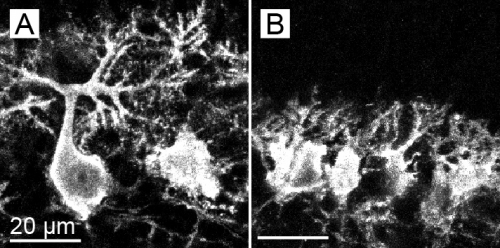
Figure 1.
Defect of dendritic development of Purkinje cells in cerebellum (postnatal day 7).
(A) Highly-branched dendrites are observed in wild-type mouse Purkinje cells.
(B) Purkinje cells in Pex14ΔC/ΔC mouse show the defect of dendritic development.
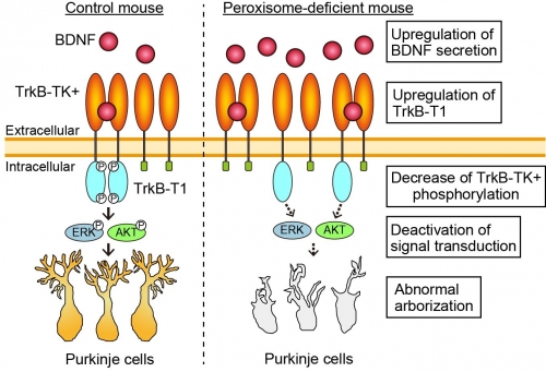
Figure 2.
In the wild-type cerebellum (left), BDNF targets to TrkB-TK+ on the surface of Purkinje cells. Cytosolic tyrosine kinase domain (TK) then undergoes autophosphorylation and activates ERK and AKT signaling, leading to the dendritic arborization of Purkinje cells. In the Pex14ΔC/ΔC mouse, BDNF level is increased around the Purkinje cells. On the Purkinje cells of Pex14ΔC/ΔC mouse (right), TrkB-T1 is upregulated and dominant-negatively inhibits autophosphorylation of TrkB-TK+. Decrease of TrkB-TK+ phosphorylation deactivates the ERK and AKT signaling, resulting in the dysmorphogenesis of Purkinje cells.
Journal Reference
Peroxisome biogenesis deficiency attenuates the BDNF-TrkB pathway-mediated development of the cerebellum, , Life Science Alliance, 10.26508/lsa.201800062.Research-related inquiries
- TOP
- News
- Research Results
- Pathological mechanism of peroxisome biogenesis disorders