![]()


![]()
![]()
研究成果 Research Results
- TOP
- News
- Research Results
- New technique reveals details of membrane structures
New technique reveals details of membrane structures
Researchers gain new insight into the packing of molecules in lipid-raft-like ordered domains using a technique called low-flux scanning electron diffraction 2021.03.03Research ResultsLife & HealthPhysics & Chemistry
Membranes responsible for numerous functions form the boundary of every single living cell, but despite advances in understanding these critical membranes, many details are still unknown.
Now, a technique developed by researchers in Kyushu University’s Faculty of Science could shed new light on how the membranes work by helping to clarify the structure of so-called lipid rafts found within them.
Acting as a platform to pass signals between the inside and outside of a cell, lipid rafts are rigid and ordered domains in the membrane made of water-insoluble molecules known as lipids.
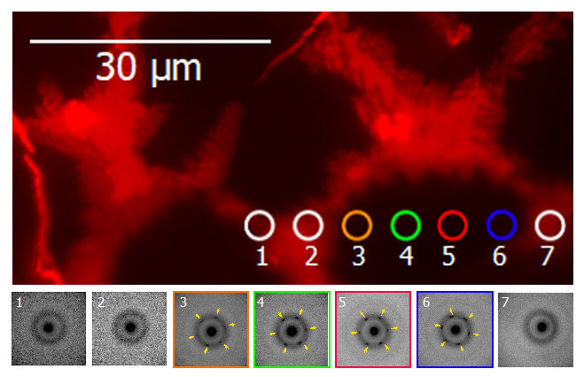
However, the packing structure of lipids inside raft-like ordered domains is poorly understood because the domains, which are often 10−30 μm in length, are much smaller than the diameter of X-rays (>100 μm) often used to probe such ordered structures, preventing the X-rays from locally examining just the tiny domains.
To overcome this problem, Nobuaki Matsumori, professor, and Masanao Kinoshita, assistant professor, at Kyushu University’s Faculty of Science, switched the X-rays with a beam of electrons having a diameter of 2.8 μm.
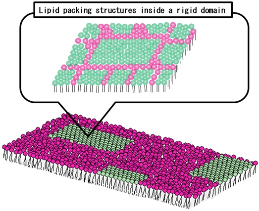
Schematic illustration of the distribution of saturated (green) and unsaturated (red) lipids in a membrane monolayer and their distribution inside the ordered domain (inset).
Using this technique, called low-flux scanning electron diffraction or LFSED for short, they studied artificial mixed membranes of saturated and unsaturated lipids that naturally separate into raft-like ordered domains within a fluid matrix.
To avoid possible damage to the membrane caused by the electron beam, the researchers reduced the number of electrons in the beam by nearly 99% compared to that used for similar experiments on other materials.
Scanning the electron beam on the raft-like ordered domains, they analyzed the bending of electrons caused by the ordered structures—a phenomenon known as diffraction—and found that individual ordered domains consist of multiple subdomains with different orientations of lipid packing.
Moreover, subdomains were found to be larger at the center of the domain and smaller near the phase boundary.
While these are just some of the first new results using the technique, the researcher expect that LFSED will be promising to investigate and develop a more detailed picture of local structures in lipid membranes.
###
For more information about this research, see “Low-flux scanning electron diffraction reveals substructures inside the ordered membrane domain,” Masanao Kinoshita, Shimpei Yamaguchi, and Nobuaki Matsumori, Scientific Reports (2020). https://doi.org/10.1038/s41598-020-79083-7
This release is also available in Japanese.
Research-related inquiries
Masanao Kinoshita, Assistant Professor
Department of Chemistry, Faculty of Science
Contact information can also be found in the full release.
- TOP
- News
- Research Results
- New technique reveals details of membrane structures