![]()


![]()
![]()
研究成果 Research Results
- TOP
- News
- Research Results
- Scanning electron microscopy technique for imaging magnetic domains expected to be applicable to many magnetic materials and devices
Scanning electron microscopy technique for imaging magnetic domains expected to be applicable to many magnetic materials and devices
2016.12.15Research ResultsPhysics & ChemistryMaterialsTechnology
Magnetic materials are important in engineering because they are used in many devices such as motors, magnetic recording media, and transformers. Generally, magnetic material is divided into small magnetic domains to lower its energy. In magnetic materials research, it is important to understand the magnetic domain structure in detail.
The magnetic domain structure can be observed easily by conventional scanning electron microscopy. However, this classic method has not been used widely because it allows observations of only limited portions of the magnetic domains, and thus only limited of magnetization components.
We have developed a visualization technique for the magnetic domain structure using an annular electron detector. We showed that the whole magnetic domain structure is quickly and easily acquired by our technique. Our technique has the advantage of simultaneously evaluating the magnetic domain structure and various microstructural properties, such as morphology, crystallographic orientation, and chemical composition. Therefore, we expect that the technique will be widely applicable to many magnetic materials and devices in both academic and engineering fields,
The results were published in Scientific Reports, which is a sister journal of the British science magazine Nature, at 10:00 a.m. GMT on November 22, 2016. This research was supported by a Grant-in-Aid for Challenging Exploratory Research (JSPS KAKENHI Grant Number JP15K14110) from the Japan Society for the Promotion of Science and Kyushu University Interdisciplinary Programs in Education and Projects in Research Development (27951).
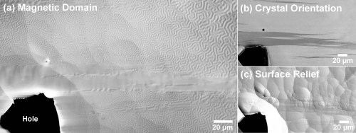
SEM images of a ferromagnetic Co-50 at.% Pt alloy. (a) A mazy domain structure is clearly visualized by the technique. (b) Crystal orientation image showing two tetragonal crystal variants with different contrasts. (c) Secondary electron image showing the surface relief caused by the Ar ion beam.
Researcher comments
Comments from the research team: SEM is a powerful tool for visualizing and analyzing a variety of microstructural properties easily over a wide area in materials. We hope that our work will expand the practical applications of SEM and advance simultaneous characterization of the structural and magnetic properties of materials.
Journal Reference
Imaging of surface spin textures on bulk crystals by scanning electron microscopy, ,Scientific Reports, 10.1038/srep37265Research-related inquiries
- TOP
- News
- Research Results
- Scanning electron microscopy technique for imaging magnetic domains expected to be applicable to many magnetic materials and devices