![]()


![]()
![]()
研究成果 Research Results
- TOP
- News
- Research Results
- Autism spectrum disorder is a “connectopathy” -Toward the development of biomarkers in the early diagnosis of ASD and early medical treatments-
Autism spectrum disorder is a “connectopathy” -Toward the development of biomarkers in the early diagnosis of ASD and early medical treatments-
2017.11.20Research ResultsLife & Health
A research group of Dr. Yamasaki and Prof. Tobimatsu of Department of Clinical Neurophysiology, Kyushu University reported the possible mechanism of atypical visual perception in autism spectrum disorder (ASD). They proposed that the“connectopathy” underlies the pathophysiological mechanism of ASD.
Serial studies on visual evoked potentials, event-related potentials, and diffusion tensor imaging conducted in our laboratory have demonstrated complex alterations (impairment and enhancement) of visual and attentional networks in ASD. Their results with a thorough paper review have suggested that the atypical visual perception observed in ASD is caused by altered connectivity within parallel visual pathways and attention networks, which contributes to the impaired social communication observed in ASD. Therefore, they suggest that the underlying pathophysiological mechanism of ASD constitutes the “connectopathy.”
This research achievement has been published on November 8, 2017, in the online edition of the Frontiers in Neuroscience.
For more information about this research, see
Yamasaki et al.: Connectopathy in autism spectrum disorders: A review of evidence from visual evoked potentials and diffusion magnetic resonance imaging.
DOI:10.3389/fnins.2017.00627
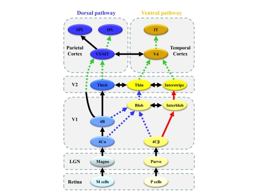
Figure 1. Schematic representation of altered connectivity of visual networks in ASD.
The human visual system is characterized by a set of parallel, hierarchical, multistage systems. There are two major parallel streams: parvocellular (or ventral) and magnocellular (or dorsal) pathways. Red thick solid arrows indicate enhanced processing (or overconnectivity), whereas blue broken arrows suggest impaired processing (or underconnectivity) in the local circuitry within the primary visual cortex (V1). Green broken arrows indicate impaired global integration of local information (or long-range underconnectivity) between lower- and higher-level visual areas.
Abbreviations: LGN, lateral geniculate nucleus; MT, middle temporal area; IPL, inferior parietal lobule; SPL, superior parietal lobule; IT, inferior temporal cortex
Journal Reference
Connectopathy in Autism Spectrum Disorders: A Review of Evidence from Visual Evoked Potentials and Diffusion Magnetic Resonance Imaging, ,Frontiers in Neuroscience, 10.3389/fnins.2017.00627Research-related inquiries
- TOP
- News
- Research Results
- Autism spectrum disorder is a “connectopathy” -Toward the development of biomarkers in the early diagnosis of ASD and early medical treatments-