![]()


-
カテゴリ別
-
場所
-
最新のお知らせ 年間スケジュール(就職対策講座等) OB・OG情報 システムの利用方法 学内合同企業説明会(「九州大学生のための業界・企業研究」) 学内企業研究セミナー / 個別企業説明会 公務員・教員等採用情報 就職相談 就職情報室 キャリア教育 低年次学年向け情報 インターンシップ等(学生向け) TOEIC対策プログラム 博士人材のための就職支援(学生向け) 外国人留学生のための就職支援 未内定の学生及び既卒者(卒業後3年以内)の皆様へ 障害のある学生のための就職支援 部局独自の就職支援 東京・大阪・博多駅オフィスの利用 各種情報サイト 過去の就職状況 採用選考に関する指針 学内合同企業説明会 博士人材のための企業説明会(企業向け) 本学へのご訪問 求人情報ご提供 OB・OG名簿ご提供 インターンシップ等(企業向け) 外国人留学生の採用 採用選考に関する指針 就職担当 よくあるご質問
Research Results 研究成果
“光生検” 切らずにその場でがんをすぐ診断
イメージングで組織を傷つけずに立体観察、AIが自動診断~ 2020.07.28研究成果Life & Health
大阪大学 大学院医学系研究科の石井優 教授(免疫細胞生物学)、松井崇浩 助教(病態病理学)、木村正 教授(産科学婦人科学)、九州大学 大学院医学研究院の加藤聖子 教授(婦人科学産科学)、株式会社ニコンの清田泰次郎氏らの研究グループは、子宮頸部を生きた組織のまま、ホルマリン固定や染色を行わずに、リアルタイムに3次元で観察できる方法を開発しました。組織の切り取りが不要なこの観察法と、人工知能(AI)による画像解析を併用することで、子宮頸がんやその超早期病変を、傷つけずに定量的に分類することができます。これらの結果は、「切り取らずに」「その場で」診断できる、新たながん診断装置の開発に役立つと期待されます。
本研究成果は、米国癌学会雑誌「Cancer Research」のオンライン版に、2020年7月23日23時(日本時間)に公開されました。
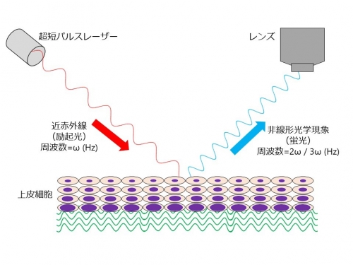
図1. 非線形光学現象による3次元蛍光イメージング
組織透過性の高い近赤外線のレーザー光を用いて、非線形光学現象で発生した蛍光を検出する。そのため、固定や染色を行っていない生きた組織でも、深部まで3次元的に観察することができる。
- 本研究についての詳細は こちら
論文情報
Nonlinear optics with near-infrared excitation enable real-time quantitative diagnosis of human cervical cancers ,Cancer Research,10.1158/0008-5472.CAN-20-0348